Understanding Autoimmunity: Causes and Healing Tips
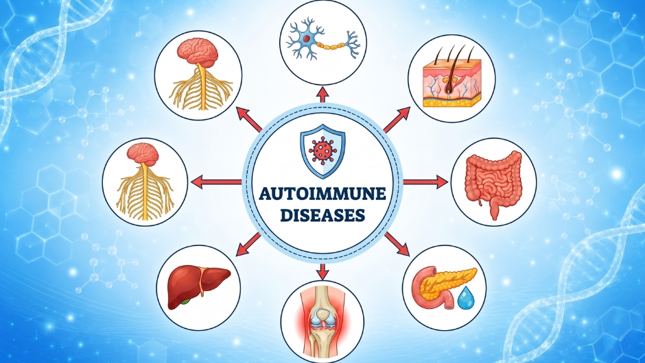
Autoimmune diseases occur when the immune system mistakenly targets self-tissues. While conventional treatment focuses on immunosuppression, a growing body of research in immunology and systems biology has identified multiple physiological factors that predispose, trigger, or perpetuate these responses. Understanding these factors does not guarantee reversal, but it provides a mechanistic roadmap for research and clinical management.
1. Increased Intestinal Permeability
The intestinal epithelium acts as a physical and immunological barrier. When tight junction proteins (e.g., occludin, claudin, zonulin) become dysregulated, luminal antigens including bacterial lipopolysaccharides (LPS) and undigested food peptides translocate into the lamina propria and circulation. This systemic antigen exposure can skew CD4+ T‑cell differentiation away from anti‑inflammatory Tregs toward pro‑inflammatory Th17 and Th1 subsets.
Note: Approximately 70–80% of immune cells reside in gut‑associated lymphoid tissue (GALT). Intestinal barrier dysfunction is observed in celiac disease, inflammatory bowel disease, type 1 diabetes, and multiple sclerosis, though causality remains an active area of investigation.
2. Gut Dysbiosis
The gut microbiome modulates immune tolerance through metabolite production. Butyrate a short‑chain fatty acid produced primarily by Firmicutes (e.g., Faecalibacterium prausnitzii, Eubacterium rectale) promotes Treg differentiation via GPR41/43 receptor signaling and histone deacetylase (HDAC) inhibition. Low butyrate levels are associated with reduced Treg function and exaggerated Th17 responses.
Dysbiosis (reduced microbial diversity, loss of butyrate producers) has been documented in rheumatoid arthritis, systemic lupus erythematosus (SLE), and ankylosing spondylitis.
3. Chronic Stress & Cortisol Dysregulation
Acute glucocorticoid elevation is anti‑inflammatory. However, chronic psychological stress leads to glucocorticoid resistance in immune cells (reduced GR‑α expression) and altered diurnal cortisol rhythms. Under these conditions, cortisol fails to suppress NF‑κB, and instead, stress‑induced catecholamines (adrenaline, noradrenaline) can promote IL‑6 and TNF‑α release.
Furthermore, chronic sympathetic activation causes microvascular damage, releasing damage‑associated molecular patterns (DAMPs) such as HMGB1, ATP, and S100 proteins, which perpetuate sterile inflammation a hallmark of autoimmune flares.
4. Estrogen Dominance
Approximately 80% of autoimmune patients are female. Estrogen metabolism influences immune cell signaling through estrogen receptors α and β expressed on lymphocytes, dendritic cells, and macrophages. At high concentrations (or with certain metabolites like 16α‑hydroxyestrone), estrogen promotes B‑cell survival, autoantibody production (e.g., anti‑dsDNA in SLE), and Th17 differentiation.
In multiple sclerosis, estrogen may have protective effects via Th2 skewing, illustrating context dependence. The key is homeostasis both estrogen excess and deficiency can be problematic depending on disease phenotype.
5. Mitochondrial Dysfunction
Mitochondria regulate immune metabolism and apoptosis. When oxidative phosphorylation is impaired (due to toxins, nutrient deficits, or genetic variants), immune cells shift toward aerobic glycolysis (Warburg effect). This metabolic reprogramming:
- Increases mitochondrial ROS (mtROS), which activate NLRP3 inflammasomes.
- Reduces ATP availability for apoptosis of autoreactive clones.
- Promotes release of mitochondrial DNA (mtDNA) as a DAMP.
Mitochondrial dysfunction has been documented in SLE, rheumatoid arthritis, primary biliary cholangitis, and type 1 diabetes.
6. Low Vagus Nerve Tone
The vagus nerve (cranial nerve X) is the primary parasympathetic efferent pathway to thoracic and abdominal viscera. It terminates at the cholinergic anti‑inflammatory pathway: acetylcholine released from vagal endings binds to α7 nicotinic receptors on macrophages and other immune cells, suppressing NF‑κB and reducing TNF, IL‑1β, and IL‑18.
Low vagal tone (measured by reduced heart rate variability, HRV) is associated with higher inflammatory markers and worse outcomes in rheumatoid arthritis and inflammatory bowel disease. Vagal nerve stimulation is an FDA‑approved therapy for some autoimmune and inflammatory conditions.
7. Nutrient Deficiencies
Immune cell differentiation from naive T cells to effector or regulatory subsets requires rapid proliferation and epigenetic remodeling. Key micronutrients involved:
- Zinc – required for thymulin activity and T‑cell maturation; deficiency impairs Treg development.
- Vitamin A (retinoic acid) – promotes gut‑homing of Tregs and inhibits Th17 differentiation.
- Vitamin D – downregulates Th1/Th17 responses and upregulates Tregs via the VDR/RXR complex.
- Vitamin C – supports epigenetic demethylation in Tregs and is an antioxidant that spares glutathione.
Without adequate levels, immune cells may remain locked in a pro‑inflammatory, undifferentiated state.
8. Omega‑6 / Omega‑3 Imbalance
Omega‑6 fatty acids (arachidonic acid, from linoleic acid in seed oils, pork, chicken) are substrates for pro‑inflammatory eicosanoids (prostaglandin E₂, leukotriene B₄) via COX and LOX pathways.
Omega‑3 fatty acids (EPA and DHA from seafood, grass‑fed ruminants) are substrates for specialised pro‑resolving mediators (SPMs), resolvins, protectins, maresins which actively terminate inflammation by promoting macrophage efferocytosis and clearing chemokine gradients.
The two classes compete for the same desaturase and elongase enzymes. A high omega‑6:omega‑3 ratio (>10:1) favours chronic, unresolved inflammation, whereas ratios near 2–4:1 are associated with lower inflammatory markers.
9. Lymphatic Insufficiency
The lymphatic system drains interstitial fluid, returns lymphocytes to the circulation, and presents antigens to lymph nodes. Effective lymphatic function requires rhythmic contraction of lymphatic vessels (powered by smooth muscle and skeletal muscle movement). When flow is compromised due to surgery, inactivity, fibrosis, or genetic lymphedema protein‑rich fluid, cellular debris, and DAMPs accumulate.
This pro‑inflammatory milieu can promote ectopic lymphoid follicle formation in target organs (e.g., thyroid in Hashimoto’s, salivary glands in Sjögren’s). Mechanistically, stasis impairs immune cell maturation and increases exposure of self‑antigens to naive T cells without proper regulatory control.
10. Oxidative Stress & NF‑κB Activation
Reactive oxygen species (ROS) superoxide, hydrogen peroxide, hydroxyl radical are generated by mitochondria, NADPH oxidases, and xanthine oxidase. At low levels, ROS serve as signaling molecules. At high or sustained levels, they directly oxidize DNA, lipids, and proteins, and crucially, activate the transcription factor NF‑κB.
NF‑κB translocates to the nucleus and upregulates hundreds of pro‑inflammatory genes: TNF‑α, IL‑6, IL‑1β, COX‑2, iNOS, and chemokines (MCP‑1, IL‑8). In autoimmunity, this pathway is constitutively active. ROS also promote protein citrullination (generating neo‑autoantigens in rheumatoid arthritis) and NETosis (neutrophil extracellular traps) in SLE.